Rutgers Scientists Reveal New Evidence of Key Mechanism in Alzheimer’s
A novel study involving human brain cells grown in mice provides insight that could lead to potential therapy
Rutgers scientists have found more clear-cut evidence of how the destructive proteins linked to Alzheimer’s disease attack human brain cells and destroy surrounding tissue.
In one of the first studies of its kind examining human brain cells grown in a mouse brain, researchers identified a pivotal mechanism that could be a potential therapeutic target for a disease that afflicts millions and has no known cure.
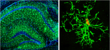
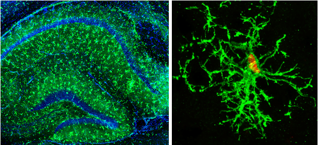
Writing in the journal Cell Stem Cell, the researchers described experiments studying human brain immune cells injected into the brains of specially bred immunodeficient mice, creating what they called a human-mouse chimera. They detailed what happened to specialized immune brain cells known as microglia after those cells were exposed to tau proteins – destructive substances believed to be involved in Alzheimer’s and other severe human brain diseases.
“In this study, we used our newly developed chimeric mouse brain model – where human cells are injected and allowed to grow, develop and mature with appropriate functions in a live mouse brain,” said Peng Jiang, an associate professor in the Department of Cell Biology and Neuroscience at the Rutgers School of Arts and Sciences. “This provided an unprecedented opportunity to investigate the role of human microglia in brains as well as the cognitive impairment seen in Alzheimer’s disease and Down syndrome, a genetic disorder with a high risk of developing Alzheimer’s disease.”
By studying the process in the human-mouse brain chimera, the scientists were able to witness and analyze – through samples extracted at different stages – a cellular brain attack that has been largely elusive up to this point.
In autopsies, scientists have been able to study the brains of people who died from Alzheimer’s and have seen residues of tau proteins, cellular changes and some other possible causative factors. The human-mouse brain chimera has allowed the Rutgers team to extract and see human cells in the actual process of deterioration.
The mice in the study were specially bred to be immunodeficient so that they could receive implanted human cells without rejecting them due to normal immune defenses. The immunodeficient mice were injected with human microglial cells and, later, with tau proteins, which are linked to the development of the brain disease.
“Since microglial cells are one of the first cell responders when something goes wrong in the brain, we believe the changes we saw to be significant,” said Mengmeng Jin, a postdoctoral researcher in the Department of Cell Biology and Neuroscience at Rutgers and first author on the study.
A genetic analysis also showed genes involved in interferon signaling turning on during the attack, indicating an important area to target for future therapies.
Other Rutgers scientists involved in the study include professor Ronald Hart, associate professors Kelvin Kwan and Ping Xie, postdoctoral fellows Ranjie Xu and Azadeh Jadali, doctoral students Mahabub Maraj Alam, Ziyuan Ma and Sining Zhu, all of the Department of Cell Biology and Neuroscience; and associate professor Zhiping Pang, postdoctoral fellow Matteo Bernabucci and doctoral student Le Wang of the Department of Neuroscience and Cell Biology and the Child Health Institute of New Jersey at Rutgers Robert Wood Johnson Medical School. Scientists at the University of California, Irvine and Florida International University contributed to the study.


