Rutgers Scientist Creates ''Virtual Biopsy'' Device to Detect Skin Tumors
Using sound vibrations and pulses of near-infrared light, a Rutgers University scientist has developed a new “virtual biopsy” device that can quickly determine a skin lesion’s depth and potential malignancy without using a scalpel.
The ability to analyze a skin tumor non-invasively could make biopsies much less risky and distressing to patients, according to a report in Wiley Online Library. Currently, physicians who perform surgical biopsies often don’t know the extent of a lesion – and whether it will be necessary to refer the patient to a specialist for extensive tissue removal or plastic surgery – until surgery has already begun.
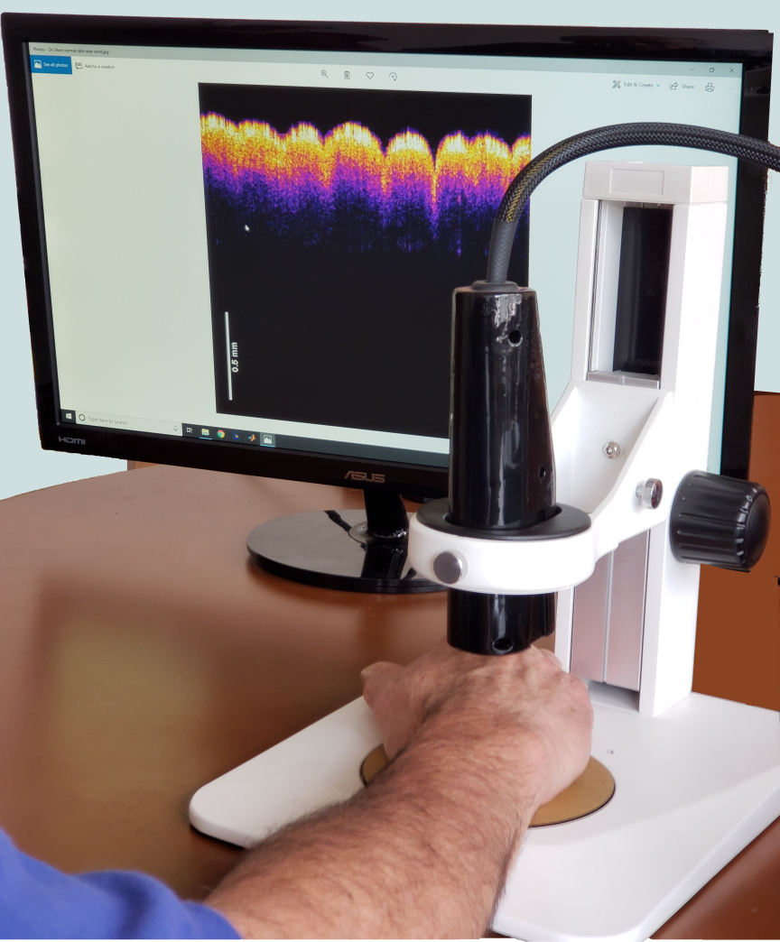
The first-of-its-kind experimental procedure, called vibrational optical coherence tomography (VOCT), creates a 3-D map of the legion’s width and depth under the skin with a tiny laser diode. It also uses soundwaves to test the lesion’s density and stiffness since cancer cells are stiffer than healthy cells. An inch-long speaker applies audible soundwaves against the skin to measure the skin’s vibrations and determine whether the lesion is malignant.
“This procedure can be completed in 15 minutes with no discomfort to the patient, who feels no sensation from the light or the nearly inaudible sound. It’s a significant improvement over surgical biopsies, which are invasive, expensive and time consuming,” said lead researcher Frederick Silver, a professor of pathology and laboratory medicine at Rutgers Robert Wood Johnson Medical School.
The study found that a prototype VOCT device, which awaits FDA approval for large-scale testing, is able to accurately distinguish between healthy skin and different types of skin lesions and carcinomas. The researchers tested the device over six months on four skin excisions and on eight volunteers without skin lesions. Further studies are needed to fine-tune the device’s ability to identify a lesion’s borders and areas of greatest density and stiffness, which would allow physicians to remove tumors with minimally invasive surgery.
Ruchit Shah, who was a Rutgers doctoral candidate at biomedical engineering at the time of the study, and researchers at the Neigel Center for Cosmetic and Laser Surgery and Center for Advanced Eye Care, co-authored the study.