Digital Cadavers Offer a High-Tech Lesson in Anatomy
"I feel like this is a tool we never knew we needed.”– Victoria Latella
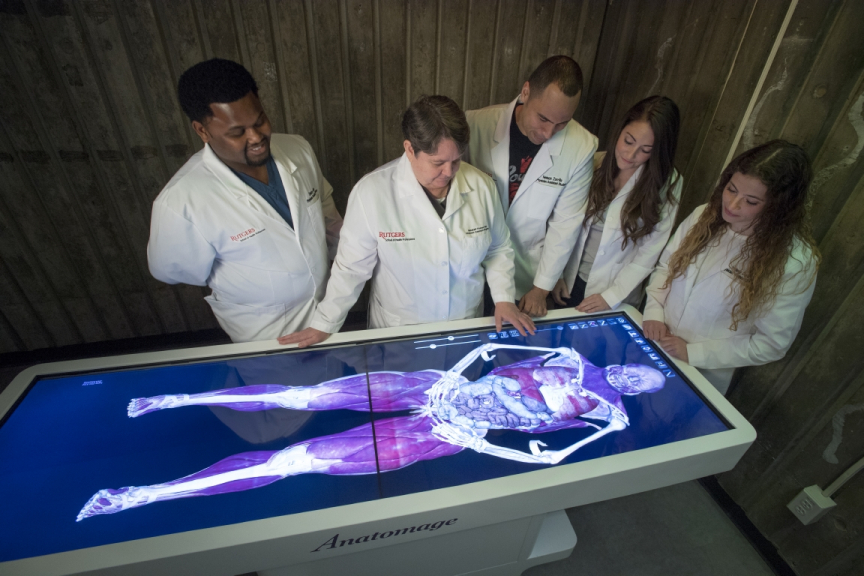
Rutgers University physician assistant students hovered over a virtual dissection table displaying the life-size image of a cadaver – the body of a 38-year-old man who had donated his body for medical research.
The minutely detailed, 3D virtual cadaver had been recreated in vivid color based on actual body scans and loaded into the 6-foot long, touch-screen table.
Swiping the screen, students peeled back layers of the cadaver, revealing pink organs and muscles, blue veins and, finally, the skeleton. To find the appendix, first-year student Lindsey DuBoff tapped on a scalpel icon, and then sliced away muscle and tissue with her finger, exposing the small organ.
Seemingly out of science fiction (it has been used by the fictional doctors on Grey’s Anatomy), the virtual dissection table brings the future of gross anatomy and clinical science education to Rutgers’ School of Health Professions, one of eight schools and clinical and education resources that comprise the university’s academic health center. Rutgers is New Jersey’s first university to use this technology.
“This table helps our students visualize and better understand anatomy and lays the foundation for stronger skills in clinical medicine,” said Matthew McQuillan, director of the physician assistant program, which confers a master of science degree.
“Many go into surgery and subspecialties and having them understand spatial relationships is critical to becoming good clinicians. Anyone doing a physical exam has to be able to visualize the body structure. The virtual dissection table helps lay a foundation to build better skills in clinical medicine.
Rebekah Thomas, an assistant professor in the program who brought the idea of the virtual cadaver to Rutgers, anticipates that it will complement – not replace – the school’s real cadavers.
Removing a kidney from a real cadaver gives students a tactile sense of the body. Students holding the organ will feel its weight and size in a way they can’t on a screen.
But a virtual kidney enables them to study the histology of an organ right down to its microscopic tissue and cells. Students can zoom in and out and see blood vessels and nerves.
In addition, once an organ is removed, a flesh-and-bone cadaver is no longer pristine. After a year of training its students with a cadaver, the university must obtain a new donor. Virtual cadavers, on the other hand, can be digitally refreshed without limit.
The computerized table gives students the flexibility to work on a variety of patient donors with different clinical and pathological conditions, body types, ethnicities and causes of death. Students also have access to a library of more than 1,000 images involving living clinical cases, which include conditions such as an ectopic pregnancy and conjoined twins – with all patients giving prior consent to the digital use of their data, according to Anatomage, the California-based company that developed the table.
“I’ve done cadaver dissections before,” said PA student Victoria Latella. “But I feel like this is a tool we never knew we needed.”